 |
||||||||||
|
|
||||||||||
|
A pioneer in glaucoma research, Anderson has pursued these questions throughout a highly productive career at Bascom Palmer. Within three years after specialty training, he was disproving assumptions widely held among ophthalmologists treating glaucoma. Indeed, pressure was a primary factor, his studies determined, but it was not the only factor. “Now we understand that it’s a spectrum,” says Anderson, who holds the School of Medicine’s Douglas Anderson Distinguished Chair in Ophthalmology. “In other words, there’s something going on in the optic nerve that makes a person susceptible to different levels of pressure. And that gradation extends down into the normal range.” Finding out how pressure causes damage to the optic nerve is the subject of current studies at Bascom Palmer, their foundations laid by Anderson’s early work that determined pressure’s role in blocking chemical messages transported along optic nerve fibers. Axons are the casualties of this blockage. Damage to the axons results in death of the parent nerve cell, which cannot be regenerated once lost. This slow but relentless cell death gradually begins to limit the range of the visual field. A constant flow of fluid that nourishes tissues in the eye naturally drains at the conjunction of the cornea and iris, called the angle. The most common type of glaucoma, known as primary open angle, occurs when this drainage channel does not do its job. As the fluid fails to properly percolate out of the eye through the angle, pressure builds. That force pushes the retina back, and its exertion on the optic nerve damages vital axons and kills the parent nerve cells. Studying these mechanisms known to create high intraocular pressure, Fini and a colleague made an important discovery, winning the New York Academy of Medicine’s 2002 Lewis Rudin Glaucoma Prize for their efforts. A tiny molecule called ELAM-1 has a tremendous effect on one of the human body’s most important functions. Fini and Joel Schuman, M.D., vice chairman of ophthalmology at Tufts University School of Medicine, spotted ELAM-1 in the tissues of patients with glaucoma. This breakthrough marked the first findings of a molecular marker for glaucoma. Responding to stress in the eye, the body produces ELAM-1, which is known to attract white blood cells to protect against infection in the vascular system. Fini and Schuman found for the first time in the eye that the stress response serves to protect cells and tissues in the pathways that regulate the critical outflow of fluid. However, they postulate that chronic activation of this response ultimately turns bad. In follow-up studies with Schuman and new collaborators at Bascom Palmer, Fini is looking for genetic modifications that could increase the deleterious effects of stress response activation, increasing a person’s chances of developing glaucoma. “The stress response in the eye’s aqueous outflow pathways is absolutely connected with glaucoma,” she says. “We believe that it will be possible to identify a genetic profile to predict a person’s risk for developing the disease, allowing us to tell if people will develop glaucoma before they get it.” “The word ‘glaucoma’ stirs up great emotional feelings,” he says. “It creates an impression before you actually get beyond that to what it really means.” The tough part is that damage to the optic nerve from glaucoma can’t yet be prevented. Usually the disease is asymptomatic until vision begins to deteriorate through loss of periphery in the visual field. But once it’s caught and a diagnosis made, treatment can prevent it from going further. Targeting intraocular pressure seems to be the most effective noninvasive method. The Ocular Hypertension Treatment Study (OHTS), a national effort cochaired by Parrish, looked to catch glaucoma triggers in the act and offer prevention options. More than 1,500 subjects 40 to 80 years of age who exhibited elevated eye pressure but no signs of glaucoma were assigned either daily doses of eye drops that reduced pressure by about 20 percent or observation without medication. Treatment with the drops was found to reduce the development of primary open-angle glaucoma by more than 50 percent. In addition, the study identified important risk factors associated with glaucoma. “As a result of this study, we now know how to better profile the treatment of patients who have elevated pressure,” Parrish says. “Appearance of the optic nerve and corneal thickness are two important factors to consider.” Remember, though, that high pressure is not always an indicator. Harking back to his discovery that susceptibility to various levels of pressure is the true culprit, Anderson and colleagues are now completing the Normal Tension Glaucoma Study (NTGS), a 15-year effort involving more than 20 centers around the world. Its results are offering solutions to clinicians who were baffled by glaucoma’s effects on patients with intraocular pressure readings falling short of the danger zone. The answer: lower the pressure regardless. “Indeed, the study group members who had their pressure lowered fared better. So pressure is involved, even if it starts at a normal range,” Anderson says. “All of our research is aimed at trying to understand the disease,” Anderson says. “I think we’re coming to understand that, among many other important factors, not being able to regulate the flow of blood sufficiently means a risk of some harm.” Bascom Palmer’s efforts don’t stop there. A talented team of bioengineers and physicists are analyzing and improving innovative diagnostic tools. Scanning Laser Polarimetry gives clinicians a view of the retinal nerve fiber layer for a closer look at glaucoma’s damage. Bascom Palmer’s own leader, Chairman Carmen Puliafito, M.D., M.B.A., coinvented a technique called Optical Coherence Tomography, which provides a cross-sectional image of the nerve fiber layer. Research conducted by Robert Knighton, Ph.D., and David Greenfield, M.D., is perfecting these technologies by accommodating for variations among individuals. Moving beyond the clinic, ophthalmology faculty members routinely conduct training and sponsor scientific conferences around the world. Back at home, the School of Medicine opens its doors to visiting fellows who travel from other countries to observe clinicians at an institute that has been rated one of the country’s top two ophthalmic centers for more than ten years running by U.S. News & World Report. “Our researchers feel a strong sense of urgency to discover new treatments for glaucoma,” Puliafito says. “With our commitment to building a team of the very finest researchers, physicians, and educators, Bascom Palmer will lead the way in providing the most advanced clinical care.” |
||||||||||
|
|
||||||||||
|
Deborah Phillips (B.S. ’98)
is an editor in the Office of Communication at the School of Medicine. |
||||||||||
|
|
||||||||||
|
|
||||||||||
 look
through the eyes of elders may grant timeless wisdom, a family’s
history, even a view of the world before technology ruled the day.
A closer look could find that perspective forever altered, limited
to tunnel vision as a result of eyesight loss associated with glaucoma.
look
through the eyes of elders may grant timeless wisdom, a family’s
history, even a view of the world before technology ruled the day.
A closer look could find that perspective forever altered, limited
to tunnel vision as a result of eyesight loss associated with glaucoma. 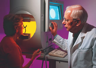
 “As you lose these cells, you lose the all-important
connection to the brain that allows you to perceive the image formed
on the retina. Once
enough cells are gone, you can’t see anymore,” says M.
Elizabeth Fini, Ph.D., who holds the Walter G. Ross Chair of Ophthalmic
Research and serves as scientific director for the McKnight Vision
Research Center. “This tiny little tissue in the back of the
eye is killed, and that’s it. You can’t see.” Fini
wants to know how to prevent the death of those cells and what’s
causing the pressure that’s behind it.
“As you lose these cells, you lose the all-important
connection to the brain that allows you to perceive the image formed
on the retina. Once
enough cells are gone, you can’t see anymore,” says M.
Elizabeth Fini, Ph.D., who holds the Walter G. Ross Chair of Ophthalmic
Research and serves as scientific director for the McKnight Vision
Research Center. “This tiny little tissue in the back of the
eye is killed, and that’s it. You can’t see.” Fini
wants to know how to prevent the death of those cells and what’s
causing the pressure that’s behind it.