 |
 |
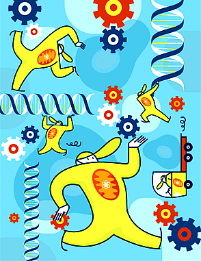
![]()
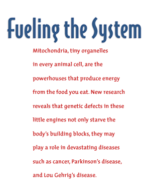
hink of them as gas-guzzlers. They are the systems
in your body that require the most fuel to function at high performance:
you r heart, your
eyes, your central nervous system. In a healthy human body, the supply
meets the demand. But in some, when the functions of tiny cellular
components called mitochondria hit a roadblock, the result is an energy
crisis.
And the systems that require the highest levels of energy are the hardest
hit.
Like the components of a well-oiled machine, the cells in your body convert the food you eat into energy. The primary role of mitochondria is to assemble specific proteins that prompt a biological chain of events called oxidative phosphorylation, which metabolizes the nutrients we ingest. At the end of the line, energy in the form of the chemical adenosine triphosphate, or ATP, is produced. Cells use this energy to do their jobs and keep us healthy.
Among the multitude of cellular components that sustain biological function, mitochondria—500 to 2,000 of which are found in each cell—are made up of their own complex machinery. In some people, mutations in mitochondria cause a breakdown. Without enough energy to fuel the body’s biological systems, the consequences can be fatal.
Beyond the primary result of an energy deficiency, there are a growing array of diseases, ranging from Parkinson’s disease to cancer, believed to be linked to mitochondrial malfunction. Focusing on the mutations that can cause such disorders, School of Medicine researchers are applying novel techniques to better understand the direct and indirect roles of mitochondria and what happens when something goes wrong.
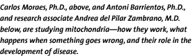
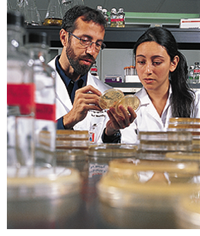
“Mitochondria are the powerhouses of the cell,” says Carlos T. Moraes, Ph.D., associate professor of neurology and cell biology and anatomy. “From the cell biologist’s point of view, the mitochondrion is a beautiful organelle. It’s really a large constellation of aspects to study.”
Defined at the genetic level for the first time in 1988, mitochondrial disorders still remain a mystery of sorts. With symptoms ranging from blindness to seizures to muscle weakness, sometimes all within the same patient, a diagnosis can be exceedingly difficult for clinicians to pin down. A slew of syndromes can present at birth, childhood, adolescence, and early adulthood—depending on the particular genetic mutation, which can be inherited from both parents or only a mother’s DNA. In some cases the mutation is not hereditary, appearing sporadically during embryo development. Scientists studying mitochondrial dysfunction investigate a host of cellular processes, including protein production, intracellular transport, even the interaction between DNA from two different sources—the nucleus and the mito-chondria. This interaction helps form the machinery that ultimately churns out energy (ATP).
![]()
tochondrial diseases were originally
identified as muscular diseases. Over time, we’ve found that
more and more disorders are due to abnormalities in mitochondrial function,” says
Walter G. Bradley, D.M., chairman of the Department of Neurology, who
was instrumental in recruiting Moraes and other innovators to boost
the school’s mitochondrial research efforts. “There is
no question that these studies enhance our neuroscience research. Our
increased ability to advance science in this area of human disease
is bringing about real breakthroughs and understanding.”
 |
||
 |
||
In the mitochondria, oxidative phosphorylation and a specific assembly of proteins called the respiratory chain allow for the breakdown of nutrients to produce ATP. If a step along this chain is halted because of a mutation in one of the necessary elements, not only is there a lack of energy, but the resulting backup can produce excessive amounts of charged molecules, also called free radicals, which are natural byproducts of mitochondrial respiration that can be harmful when overproduced.
“A problem in the respiratory chain increases the creation of free radicals that may damage cells that are more susceptible, such as neurons,” says Moraes. “This puts mitochondrial dysfunction in a group much larger than mitochondrial diseases because it can extend to neuro-degenerative diseases.”
With so much to investigate, where is the starting point for correcting or preventing mitochondrial dysfunction? Scientists at the School of Medicine are heading back to the basics, constructing their studies with the basic building blocks of life.
“If you define life as the ability of cells to divide and replicate, DNA is the source of life,” says Louis Elsas, M.D., director of the school’s Dr. John T. Macdonald Foundation Center for Medical Genetics. “The double helix structure of DNA allows for replication and change in the transmission of characteristics from generation to generation. DNA is located in the nucleus and in the mitochondria, which also provide the energy necessary for DNA to divide and replicate.”
 Housed in the nucleus of each of our body’s cells are the DNA
strands that form the genes to make us the individuals we are. Along
with establishing characteristics such as hair color or the accuracy
of your eyesight, nuclear genes are responsible for coding particular
proteins and sending them to the mitochondria for their place in the
oxidative phosphorylation chain. They are joined in the process by
proteins encoded by DNA located exclusively in the mitochondria. Malfunctions
in the mitochondria result from malfunctions in the coded proteins,
which brings the blame back to the cell’s genes (both nuclear
and mitochondrial DNA can mutate). The answers, therefore, must lie
in the human genome.
Housed in the nucleus of each of our body’s cells are the DNA
strands that form the genes to make us the individuals we are. Along
with establishing characteristics such as hair color or the accuracy
of your eyesight, nuclear genes are responsible for coding particular
proteins and sending them to the mitochondria for their place in the
oxidative phosphorylation chain. They are joined in the process by
proteins encoded by DNA located exclusively in the mitochondria. Malfunctions
in the mitochondria result from malfunctions in the coded proteins,
which brings the blame back to the cell’s genes (both nuclear
and mitochondrial DNA can mutate). The answers, therefore, must lie
in the human genome.
Antoni Barrientos, Ph.D., assistant professor of neurology at the Dr. John T. Macdonald Foundation Center for Medical Genetics, is searching for those answers in a seemingly unlikely source. Not just handy in the bakery, yeast is proving to be an optimal model for studying the human genome.“There is little difference between human and yeast mitochondrial proteins involved in the production of energy,” Barrientos says. “The genes related to energy production have been conserved through evolution.” The model organism also is easy to replicate and maintain in the lab. And with the entire yeast genome sequenced since 1996, joined by the human genome more recently, Barrientos and his colleagues are focusing now on functional genomics, defining the roles of the genes various organisms share and how the proteins they encode relate to mitochondrial function.

“ If you look at evolutional scales—where genes have been conserved across the genomes of different organisms—you can theorize that these are important genes,” says Elsas. “And if they are altered, some major deficiency should occur. Then we can ask, ‘How does that mutation affect other genes and their functions? What are the links between these pathways? How do they interface?’” These questions have resulted in a field of genetics called “metabolomics” or systems biology.
To find out if a mutation in a particular gene will cause mitochondrial malfunction, Barrientos is creating “knockout” yeast models, in which he removes the normal gene and replaces it with a mutated version. Based on tests such as observing the yeast cell’s subsequent growth or measuring the cell’s consumption of oxygen, Barrientos classifies the particular genetic mutation as either harmful to energy production or inconsequential.
The next step is studying the potential of these discoveries for creating clinical treatments. In Moraes’s lab, researchers target mutated genes by introducing proteins that bind to and digest the mutated strands of DNA, ridding the cell of these culprits of disease. The cell responds by creating more of the existing healthy DNA to recharge its genetic supply. While still far from application in the clinic, these studies lay important groundwork and have received recognition in journals such as Human Molecular Genetics.
Another promising avenue of study is the genetic interaction between the DNA of the nucleus and DNA unique to the cell’s mitochondria. Since both are important players in the respiratory chain to produce energy, Moraes wants to know how much mutation in either set of DNA is too much; for example, what is the threshold at which dysfunction occurs? By mixing the mitochondrial DNA from one species with the nuclear DNA of another, he is able to identify the variability allowable for mitochondria to continue normal function. So far, studies have determined that even small changes make a big difference. Human DNA, for example, is compatible only with that of species who are close genetic kin, such as chimpanzees. Beyond such a close match, mitochondrial function cannot proceed as normal.
“This tells us that interaction between nuclear DNA and mitochondrial DNA is very precise and sensitive to evolutionary development,” Moraes says.
Researchers also are studying the role of mitochondria in cell death. Collaborating with Lawrence Boise, Ph.D., associate professor at the University of Miami Sylvester Comprehensive Cancer Center, Moraes is focusing on how mitochondria may contribute to apoptosis, the body’s natural renewal process that stimulates cell death. In a normal functioning cell, proteins in mitochondria control the release of cytochrome c, an electron transport molecule that transmits a signal to begin apoptosis. But when too much cytochrome c is released, apoptosis may spiral out of control, as is the case in the loss of neurons associated with Parkinson’s disease. When too little cytochrome c is released, the reverse happens and natural cell death is not allowed, setting the stage for uncontrolled growth of malignant or abnormal cells. Since the signal for mitochondria to release cytochrome c begins outside the organelle, Moraes and Boise have much to study in determining where communication of apoptosis goes awry.
“This research spans so many different departments,” says Bradley. “We have a sign here that says, ‘collaborate or die.’ It really is the most important thing.” Faculty from a range of departments, including immunology, physiology, neurology, pathology, cell biology, pharmacology, and others are given the opportunity for such collaboration at monthly meetings of the interdisciplinary Mitochondria Research Club. Presentations and discussions give these scientific minds a creative outlet that often has led to innovative approaches across disciplines.
Moraes, who coordinates the club, says, “Sometimes the best ideas come from something that’s related to but not exactly what you’re studying. It gives you a new perspective.”
“Meeting with clinical and basic resear-chers is like bringing together two different worlds,” adds Barrientos. “But these kinds of interactions help us focus and also give us validation of the work we’re doing.” According to Norman Altman, V.M.D., vice provost for research, further validation has come from an increase in federal grants to the School of Medicine to study mitochondrial function.
As with every laboratory pursuit, the answer to one question leads to many others. School of Medicine researchers find themselves on a track toward discovery and ultimately treatment for patients suffering from mysterious and often devastating disorders.