 If given in high enough doses, you can kill anything with radiation—except, as I understand, some types of Florida cockroaches. There is no question that radiation is an effective local treatment for cancer. In radiation oncology, the conventional wisdom is to administer as high a dose as necessary to completely eradicate the cancer while sparing the surrounding normal tissues. If given in high enough doses, you can kill anything with radiation—except, as I understand, some types of Florida cockroaches. There is no question that radiation is an effective local treatment for cancer. In radiation oncology, the conventional wisdom is to administer as high a dose as necessary to completely eradicate the cancer while sparing the surrounding normal tissues.
Although this strategy is straight- forward, tumors have a habit of locating themselves next to vital or sensitive normal structures, and they often move during treatment. There are several methods that have recently come into practice that are designed to deal with these problems.
 The field of radiation oncology has changed so dramatically over the last 20 years that even some of the fundamental tenets of the specialty have been challenged. The development of stereotactic radiosurgery/radiotherapy is a prime example. The precision of radiation delivery has improved so much that we can administer very high doses to small volumes and eliminate a tumor in one to five treatments, instead of 20 to 35. From lung to liver to brain cancer, great advances have been made using stereotactic radiotherapy, and the future looks even brighter. At the University of Miami, we are investing heavily in state-of-the-art equipment for a Stereotactic Radiosurgery Center of Excellence. Our team of investigators will integrate these techniques with novel methods of tracking tumors in 4D and sophisticated functional imaging methods. The field of radiation oncology has changed so dramatically over the last 20 years that even some of the fundamental tenets of the specialty have been challenged. The development of stereotactic radiosurgery/radiotherapy is a prime example. The precision of radiation delivery has improved so much that we can administer very high doses to small volumes and eliminate a tumor in one to five treatments, instead of 20 to 35. From lung to liver to brain cancer, great advances have been made using stereotactic radiotherapy, and the future looks even brighter. At the University of Miami, we are investing heavily in state-of-the-art equipment for a Stereotactic Radiosurgery Center of Excellence. Our team of investigators will integrate these techniques with novel methods of tracking tumors in 4D and sophisticated functional imaging methods.
The tactic of concurrent radiation with chemotherapy has resulted in greater cure rates for many cancers, including lung, head and neck, gastrointestinal, and cervical cancers. We have slowly learned how to combine radiation with chemotherapy, but the price has been high in terms of side effects.
The dream of individualized cancer care is becoming a reality—patients are being assessed for gene expression abnormalities (biomarkers) more frequently to determine the most appropriate treatment. Gene expression profiles, along with the person’s genetic makeup (DNA sequence), will be used to predict response and complication risk to radiation and agents that are combined with radiation. In collaboration with other investigators at the University of Miami and at a number of other academic centers around the country, there is a major ongoing research effort in the Department of Radiation Oncology to bring newly identified biomarkers that predict outcomes in prostate cancer patients treated with radiotherapy into more routine clinical practice.
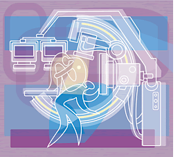
The most common method of administering radiation to treat cancer is external beam radiotherapy, using X-rays that are photons. Sprouting up across the United States are facilities, costing $120 million and more, designed to accelerate and deliver proton particle therapy. The advantage of protons is they travel a certain distance in the body and abruptly stop. When they stop, they deposit a large bolus of energy called the Bragg peak, which can be placed at the site of a tumor deep in the body. Whereas photons continue through the body, protons have much less of an exit dose.
The University of Miami is investing in proton therapy because the technology is tremendously promising, and for some cancers the promise is evident today. The future of radiation oncology rests on the development of modalities that improve normal-tissue sparing (e.g., proton therapy), the application of functional imaging to better identify the tumor, and improvements in tumor tracking techniques during treatment. Advancements in these areas, combined with tumor gene expression profiling to select patients and the addition of targeted radiotherapy, will lead to much higher cure rates. The University of Miami is well positioned to be at the forefront of this new age in radiation oncology.
Alan Pollack, M.D. ’87, Ph.D. ’79, was named chair of the Department of Radiation Oncology last year.
|