|
|
Revealing Images
By Dwayne Campbell | Photos by Byron Maldonado and John Zillioux
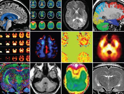
New techniques in magnetic resonance
and other imaging technologies being pioneered by Miller School faculty are paving the way for more accurate evaluation of several complex conditions.
Richenel Jean-Pierre has almost no memory of the crash. When he regained consciousness in the hospital, his family, on the advice of doctors, gave him few details. They would not tell him his car was totaled. They would not tell him he had come face-to-face with death.
Two years later it is difficult to tell that Jean-Pierre was a victim of brain trauma. But, he says, there are moments when he knows he’s a changed man. He has short-term memory loss. In striking contrast to his once quiet personality, he sometimes talks non-stop; when he gets upset, his head gets as hot as flames. “I am lucky to be alive,” says Jean-Pierre, 24. “But I am still fighting.”
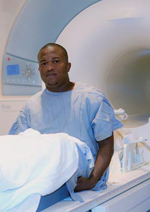 |
| Richenel Jean-Pierre, who suffered mild traumatic brain injury (TBI) in an automobile crash, is participating in a magnetic resonance imaging study designed to detect chemical changes in the brain after TBI |
Jean-Pierre is one of several people participating in a complex magnetic resonance imaging (MRI) study designed to detect changes in several chemical compounds in the brain following mild traumatic brain injury (TBI). Headed by Andrew Maudsley, Ph.D., professor of radiology and principal investigator, and Varan Govind, Ph.D., the program is among several cutting-edge MRI projects being conducted by researchers who are bringing national renown to the Miller School’s Department of Radiology and its interdisciplinary work in imaging research.
Maudsley, a pioneering researcher in new imaging techniques who trained under two Nobel Prize winners (Sir Peter Mansfield, who won the prize for physics in 2003, and Richard Ernst, who won for chemistry in 1991), moved to UM in 2002, joining notable faculty such as assistant professor Michalis Georgiou, Ph.D., an expert in nuclear medicine physics and instrumentation. Maudsley helped set the stage for recruiting other key researchers such as Govind, a research assistant professor of radiology and expert in nuclear magnetic resonance (NMR) spectroscopy who evaluates new proton (hydrogen) spectroscopic imaging techniques for diagnostic imaging applications.
“We have significantly increased our emphasis on imaging research, and to that end we have hired innovative and cutting-edge Ph.D. faculty members whose areas of concentration involve advanced imaging,” says Robert Quencer, M.D., professor and chair of radiology. “We have also developed a state-of-the-art image processing lab, which will be available to clinicians and researchers so that processing of imaging data can yield more information about various disease processes. This will all translate into better patient care.”
Rapid advancements in imaging have made radiology a focal point of modern academic medicine. For example, new developments in MR mammography have changed the way breast disease is analyzed and treated. MR imaging is now used for the evaluation of cardiac patients. Early cartilage damage in various sports injuries is analyzed with new MR software programs. And the use of MR to identify brain tissue at risk in acute stroke contributes to better survival and outcomes in these patients.
Here at the Miller School and UHealth – University of Miami Health System, researchers envision better patient care through cooperative interdisciplinary work between various labs, MR imaging, and patient care areas. “This kind of collaboration among basic scientists, computer scientists, and clinicians is what’s driving the cutting-edge developments in diagnostic imaging in most U.S. academic centers,” says Maudsley.
Some of the Miller School’s most prolific radiologic researchers and their work are profiled below.
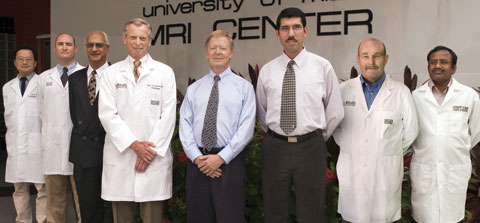 |
| Pioneering promising new clinical applications for magnetic resonance and other imaging technologies are Miller School faculty members including, from left, Jung-Jiin ‘Jason’ Hsu, Ph.D., Kyle R. Padgett, Ph.D., Pradip M. Pattany, Ph.D., Robert Quencer, M.D., Andrew Maudsley, Ph.D., Mohammad Sabati, Ph.D., Noam Alperin, Ph.D., and Varan Govind, Ph.D.
|
Andrew Maudsley, Ph.D.,
and Varan Govind, Ph.D.
Imaging Cellular Injury
The work of Maudsley and Govind has provided a unique insight into how the brain works in the aftermath of mild injury. While clinical X-ray CT and MRIs have long shed light on possible damage to brain tissue after trauma, minute changes on the cellular scale are harder to detect. Maudsley’s imaging research measures alterations in the metabolism of the tissue, looking at compounds such as N-acetylaspartate, creatine, and choline in the brain. Increased choline and decreased N-acetylaspartate are common observations in many pathologies.
“Metabolites are very difficult to evaluate by eye, so we looked for a better way to analyze subtle change in the concentration of these chemicals,” says Maudsley. “We mapped their natural distribution in the brain, and now have normative information we can incorporate into the analysis of patient studies.”
That information is important for TBI patients because there are only limited quantitative tests to assess them when they arrive at a hospital. Patients with mild injuries who experience only a brief loss of consciousness, Maudsley says, are sometimes admitted to the trauma unit but often discharged quickly. Many recover within a few weeks or months, but some continue to have problems for a longer time.
Maudsley’s and Govind’s research could provide a way to evaluate the level of injury and even indicate which patients have a high likelihood of full recovery in a given period and which may need more aggressive intervention.
Funded by the National Institutes of Health, the TBI project has been renewed for a fourth year. The potential of these imaging methods for detecting changes in brain chemistry at the millimolar level has other potential applications including brain cancer research and neurodegenerative diseases. Govind also has an NIH-funded project with collaborators in the Department of Neurology who are applying these same methods to amyotrophic lateral sclerosis (ALS).
Maudsley also has another research project, now in the fifth year of a 10-year NIH grant, to continue development of these metabolic imaging methods for use on MRI instruments across the country.
Noam Alperin, Ph.D.
MR of Vital Fluids
Noam Alperin, Ph.D., is known for his groundbreaking work in magnetic resonance imaging of flow of blood and cerebrospinal fluid (CSF) to and from the brain.
“Once we understood that CSF pulsation is driven by blood pulsation, we applied engineering methodology and control theory to study the interplay between blood and CSF flow dynamics,” says Alperin, a professor of radiology who did post-doctoral research in the area at the University of Chicago and later as a faculty member at the University of Illinois, Chicago. “We can learn a lot about pressure and compliances in the brain without making a hole in the skull.”
Alperin’s noninvasive imaging method measures important physiological parameters such as intracranial pressure and cerebral blood flow. With a $1.5 million NIH grant, the work of Alperin and his research team—Murat Bagci, Ph.D.; Sang “Aaron” Lee, M.S.; Rong-Wen Tain, M.S.; and Sudarshan Ranganathan, M.S.—could contribute to improved therapies for conditions such as hydrocephalus, idiopathic intracranial hypertension, and Chiari malformations. The team already has found predictors that can help identify patients who may benefit from brain surgery.
Alperin is also directing the creation of the Miller School’s advanced image processing lab.
Jung-Jiin ‘Jason’ Hsu, Ph.D.
MR Neuroimaging
Jung-Jiin ‘Jason’ Hsu, Ph.D., a research assistant professor of radiology who did his post-doctoral work at Stanford in functional magnetic resonance imaging, is making neuroimaging more widespread and readily available at the Miller School. He is currently working with Fatta Nahab, M.D., assistant professor of neurology, and Clinton Wright, M.D., associate professor of neurology.
Hsu is also continuing his research in functional MRI and oximetry. At Stanford, he developed a technique of longitudinal relaxation measurement that does not need the long delay time required by conventional methods, accelerating measurement sufficiently for brain functional imaging. His technique opens a new window to quantify blood perfusion and oxygenation change. He is using the technique for functional neuroimaging to explore brain areas for which conventional MRI methods generate poor image quality, as well as MRI oximetry, the measurement of oxygen concentration, which could replace current highly invasive and risky techniques that use electrodes or optical needles.
Mohammad Sabati, Ph.D.
MR Angiography
Vascular imaging is the forte of Mohammad Sabati, Ph.D., research assistant professor of radiology, whose background in electrical engineering has influenced his approach to MR imaging. Sabati’s single-acquisition method of MR angiography allows for minimal use of contrast agent materials and a continuous imaging process, even for large areas such as the lower limbs. Through the use of “continuously moving table” imaging that grew out of his Ph.D. research, Sabati captures one large, seamless image of different parts of the body, avoiding the need for multiple images and facilitating diagnosis of systemic diseases. Sabati is researching non-contrast MR angiography in, for example, patients with kidney disease who cannot tolerate contrast MRIs. He has already received a favorable review for a pending NIH grant expected to enroll 30 patients in a study he says holds hope for “avoiding contrast complications in the health care system.”
Pradip M. Pattany, Ph.D.
MR Investigations
Pradip M. Pattany, Ph.D., joined the Miller School in 1993 following stints in the MR industry, where he developed and patented MR methods in quantitative blood flow and minimizing physiologic motion. Award-winning and revolutionary at the time, the MR method allowed clear images in spite of pulsation of blood, cerebrospinal fluid flow, and respiratory or other motion. Variations of this method are now standard protocol in imaging.
Pattany, research associate professor of radiology, now concentrates on developing MR imaging methodology, including non-invasive ways that can still garner accurate results. Much of the research is dedicated to imaging spinal cord injuries, often via diffusion tensor imaging, as part of the Miller School’s interdisciplinary efforts to cure such injuries. Some of the spinal cord research uses MR to investigate neuropathic “phantom” pain and accompanying changes in certain brain metabolites.
Pattany is also using imaging in an in-vivo project to get a baseline of white matter changes during aging and imaging of the heart pre- and post-therapy.
Kyle R. Padgett, Ph.D.
Campus-Wide Collaborations
As interdisciplinary collaboration in imaging grows, more researchers are encountering Kyle R. Padgett, Ph.D., who, since 2005, has operated one of the most active MR research labs on campus. Padgett is an expert in designing appropriate protocols, monitoring the imaging process, and evaluating the results for myriad experiments across departments.
One of the most significant studies in which Padgett is involved focuses on spinal cord injury with researchers from The Miami Project to Cure Paralysis, as well as the departments of neurological surgery, pediatrics, and others. Juan Pablo Solano, M.D., assistant professor of pediatrics, says that in these complicated controlled contusion spinal cord investigations, MR is one of the best ways to assess the injury and any potential side effects of a particular treatment. In one study, MR imaging will track cell distribution after injection into an injury site to confirm that targeted cell therapy is being properly delivered and to monitor the progression of the treatment.
“MR is a powerful investigative tool, and its inclusion typically adds high scientific value to a study,” says Padgett. “MR is still an emerging field that holds tremendous promise for helping millions of patients, and UM is one of the most dynamic MR centers in the country.” |
|
|